2015 · Hemodynamics & imaging
Benefit of cone-beam CT angiography in visualizing aneurysm shape and identification of exact rupture site
Lauric A, Heller RS, Schimansky S, Malek AM.
J Neuroimaging 25(1):56-61, 2015.
PMID 24707990 · DOI 10.1111/jon.12120
In plain language
Brain aneurysms are usually imaged with standard 2D angiography or 3D rotational angiography, both X-ray studies of the blood vessels. Cone-beam CT angiography is another option that provides even finer detail.
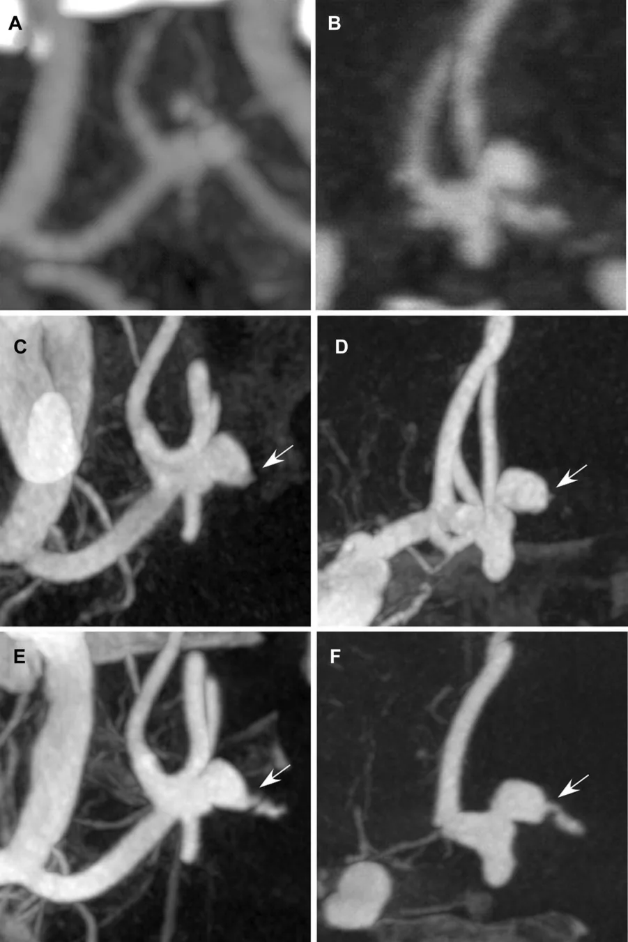
This case report describes a 54-year-old man with bleeding around the brain (subarachnoid hemorrhage) from a ruptured aneurysm on the anterior communicating artery, a small vessel connecting the left and right arteries at the front of the brain. The aneurysm's shape looked dramatically different depending on the imaging method. Only cone-beam CT clearly showed the exact rupture site, a small irregular, elongated bulge on the aneurysm dome that the other methods missed, and its detail allowed more precise computer simulations of blood flow.
Because aneurysms likely change shape before or at rupture, the authors advise that flow studies take into account the resolution of the imaging used.
A plain-language summary for patients and families. It is not medical advice, and individual results vary.
Abstract
While high-resolution cone-beam computational tomographic (CBCT) angiography has gained use in intracranial vascular imaging, digital subtraction angiography (DSA) and 3-dimensional-rotational angiography (3D-RA) remain the preferred acquisition modalities for intracranial aneurysm imaging. This case report highlights the utility of the greater spatial resolution afforded by CBCT for cerebral aneurysm imaging. A 54-year-old man presenting with subarachnoid hemorrhage was confirmed to harbor a ruptured anterior communicating artery aneurysm by conventional angiography. Due to varying contrast opacification captured by different acquisition methods, dramatic aneurysm shape difference was observed between 2- and 3-dimensional-angiographic and CBCT models. The greater resolution of CBCT revealed in an unequivocal fashion the exact site of rupture on the aneurysm dome, visualized as a discrete irregular and elongated bleb that was not seen on either 3D-RA or DSA. High-resolution CBCT visualized the shape of the target aneurysm in greater detail than the more conventional 2D-DSA and 3D-RA, enabling more precise computational fluid dynamics (CFD) simulations. Given that aneurysms most likely change shape either prior to rupture or upon rupture, future studies evaluating fluid dynamics using computer reconstructions should be cognizant of the differences in resolution provided by various imaging modalities.
Abstract as indexed in PubMed (PMID 24707990). Copyright the publisher.
Cite this paper
Lauric A, Heller RS, Schimansky S, Malek AM. Benefit of cone-beam CT angiography in visualizing aneurysm shape and identification of exact rupture site. J Neuroimaging 25(1):56-61. 2015. doi:10.1111/jon.12120 PMID: 24707990.