2014 · Hemodynamics & imaging
Benefit of a sharp computed tomography angiography reconstruction kernel for improved characterization of intracranial aneurysms
Oʼmeara B, Rahal JP, Lauric A, Malek AM.
Neurosurgery 10 Suppl 1:97-105; discussion 105, 2014.
PMID 24030173 · DOI 10.1227/NEU.0000000000000167
In plain language
CT angiography, a fast CT scan of the blood vessels, is usually the first test for brain aneurysms, but catheter angiography is often needed to see their shape in detail. CT images can be rebuilt from the raw data using different settings called kernels, some smoother and some sharper.
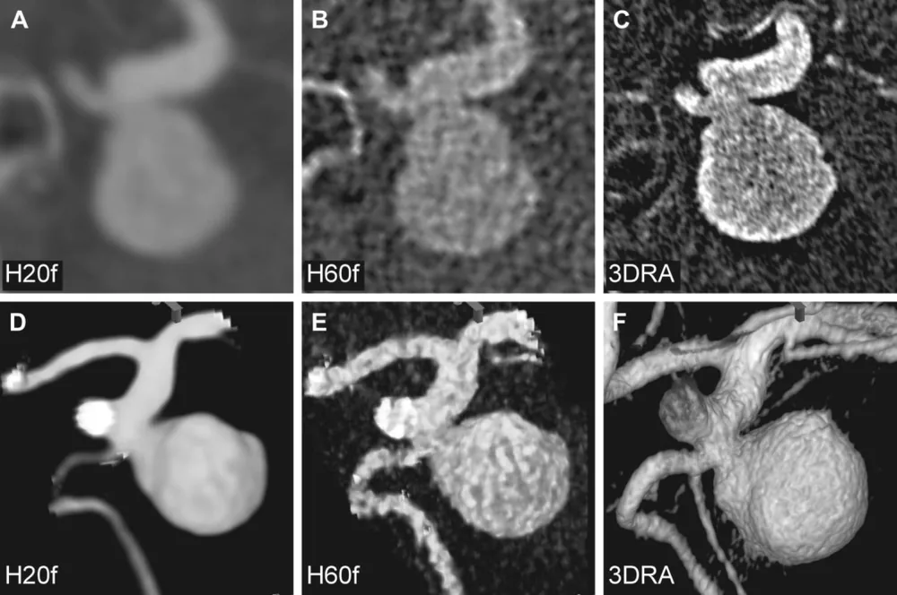
In 15 patients with aneurysm bleeding, CT images were rebuilt with a standard smooth kernel and a sharp kernel and compared with 3D catheter angiography. The smooth kernel overestimated the aneurysm neck by about 0.64 millimeters compared with the sharp kernel, while the sharp kernel matched catheter angiography with no significant difference.
The sharp setting gives more accurate measurements, though the images look less smooth. It needs no extra contrast, radiation or equipment, and may help with treatment planning.
A plain-language summary for patients and families. It is not medical advice, and individual results vary.
Abstract
Background
Computed tomography angiography (CTA) is the first-line imaging modality used for cerebral aneurysms because of its speed and sensitivity for detection, although digital subtraction angiography is often required for more detailed aneurysm shape delineation.
Objective
To determine whether a sharper CTA reconstruction kernel can better characterize an aneurysm and improve decision-making before intervention.
Methods
Fifteen patients presenting with aneurysmal subarachnoid hemorrhage underwent 64-row CTA. CTA data were reconstructed using the default H20f smooth kernel and a H60f sharp kernel and compared with contemporaneous catheter 3-dimensional rotational angiography (3DRA). Aneurysm neck, width, and aspect ratio measurements were made using intensity line plots of identical projections on all imaging datasets and compared by matched-pair statistics.
Results
Aneurysm neck measurements from the H20f smooth kernel revealed overestimation compared with both the sharp kernel (greater by 0.64 ± 0.21 mm, P < .01) and 3DRA (greater by 0.68 ± 0.19 mm, P < .01). There was no statistically significant difference between 3DRA and the sharp kernel CTA measurements. Neck measurements correlated well between the H60f kernel and 3DRA but not between the H20f Kernel and 3DRA (R 0.97 vs 0.86).
Conclusion
H60f sharp CTA kernel reconstruction provides more accurate anatomic characterization of cerebral aneurysms than the H20f smooth kernel at the expense of less visually pleasing reconstructions. Because it does not require additional contrast, radiation, or imaging hardware and is more similar to 3DRA, it may aid in selecting the appropriate treatment strategy before to evaluation by catheter-based angiography.
Abstract as indexed in PubMed (PMID 24030173). Copyright the publisher.
Cite this paper
Oʼmeara B, Rahal JP, Lauric A, Malek AM. Benefit of a sharp computed tomography angiography reconstruction kernel for improved characterization of intracranial aneurysms. Neurosurgery 10 Suppl 1:97-105; discussion 105. 2014. doi:10.1227/NEU.0000000000000167 PMID: 24030173.