2006 · Hemodynamics & imaging
Fusion of three-dimensional calcium rendering with rotational angiography to guide the treatment of a giant intracranial aneurysm: technical case report
Hoit DA, Malek AM.
Neurosurgery 58(1 Suppl):ONS-E173; discussion ONS-E173, 2006.
PMID 16462621 · DOI 10.1227/01.NEU.0000193524.98940.BB
In plain language
Three-dimensional rotational angiography produces detailed 3D pictures of brain arteries, but it shows calcium deposits poorly. Calcium near the neck of an aneurysm can affect how it is best treated.
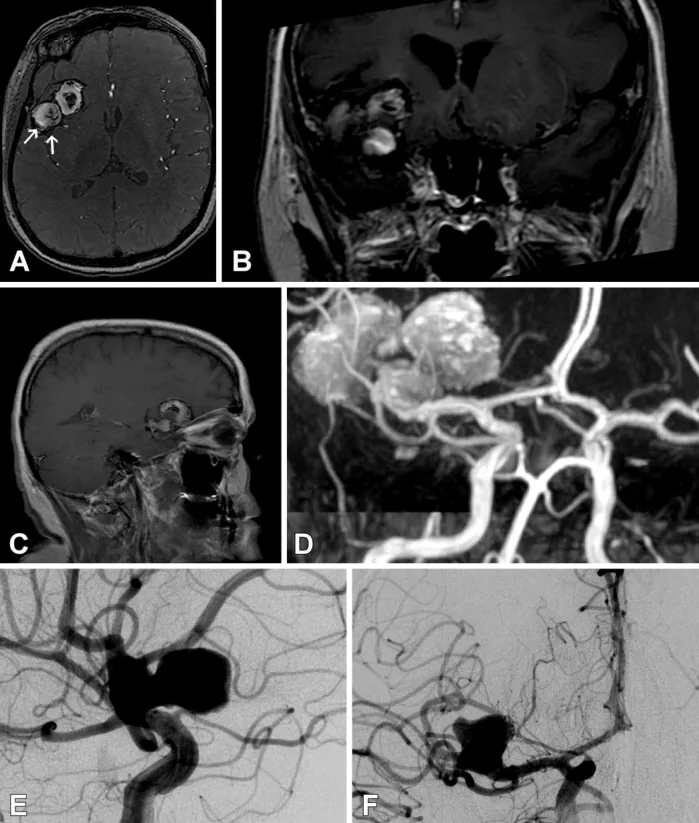
This technical report describes a 57-year-old woman with a family history of brain bleeding who was found to have a giant, calcified, partly clot-filled aneurysm of the middle cerebral artery. The team performed a separate scan tuned to show calcium and merged it with the standard 3D vessel image. The combined view revealed heavy calcium at the neck of the aneurysm that ruled out surgical clipping, so she was treated with coils from inside the vessel instead.
The method can help plan treatment by revealing calcium that would otherwise be missed.
A plain-language summary for patients and families. It is not medical advice, and individual results vary.
Abstract
Objective and importance
Although three-dimensional (3D) rotational angiography enables the visualization of intracranial vascular anatomy with high spatial resolution, its ability to resolve calcified features is limited. We present a case in which a 3D calcium density rendering was fused with rotational cerebral angiography to guide the treatment strategy of a giant, calcified, partially thrombosed, middle cerebral artery bifurcation aneurysm.
Clinical presentation
On screening magnetic resonance imaging, a 57-year-old woman with familial subarachnoid hemorrhage was found to harbor a giant trilobed, partially thrombosed, calcified aneurysm. Three-dimensional (3D) cerebral angiography revealed only partial contrast filling of one lobe with a daughter component.
Technique
A distinct rotational acquisition volume with parameters optimized for calcium density visualization was acquired just before the conventional contrast acquisition. The resulting 3D calcium volume rendering was fused with the vascular model. Multiplanar interactive visualization of the combined model uncovered heavy calcification at the neck of the aneurysm that precluded direct clipping, and the patient underwent endovascular coil embolization.
Conclusion
Preoperative assessment of intracranial aneurysms can be facilitated by the current technique, which enables the detection of calcified tissue near the neck, which would otherwise not be possible with 3D rotational cerebral angiography alone.
Abstract as indexed in PubMed (PMID 16462621). Copyright the publisher.
Cite this paper
Hoit DA, Malek AM. Fusion of three-dimensional calcium rendering with rotational angiography to guide the treatment of a giant intracranial aneurysm: technical case report. Neurosurgery 58(1 Suppl):ONS-E173; discussion ONS-E173. 2006. doi:10.1227/01.NEU.0000193524.98940.BB PMID: 16462621.