2005 · Devices & clinical
Three-dimensional rotational angiographic detection of in-stent stenosis in wide-necked aneurysms treated with a self-expanding intracranial stent
Hoit DA, Malek AM.
Neurosurgery 57(6):1228-36; discussion 1228-36, 2005.
PMID 16331171 · DOI 10.1227/01.neu.0000186026.12709.bc
In plain language
Wide-necked aneurysms, bulges in brain arteries with a broad opening, are sometimes treated by placing a soft, self-expanding stent (the Neuroform) across the opening and filling the aneurysm with coils. This study asked whether the stent changes the size of the artery over time.
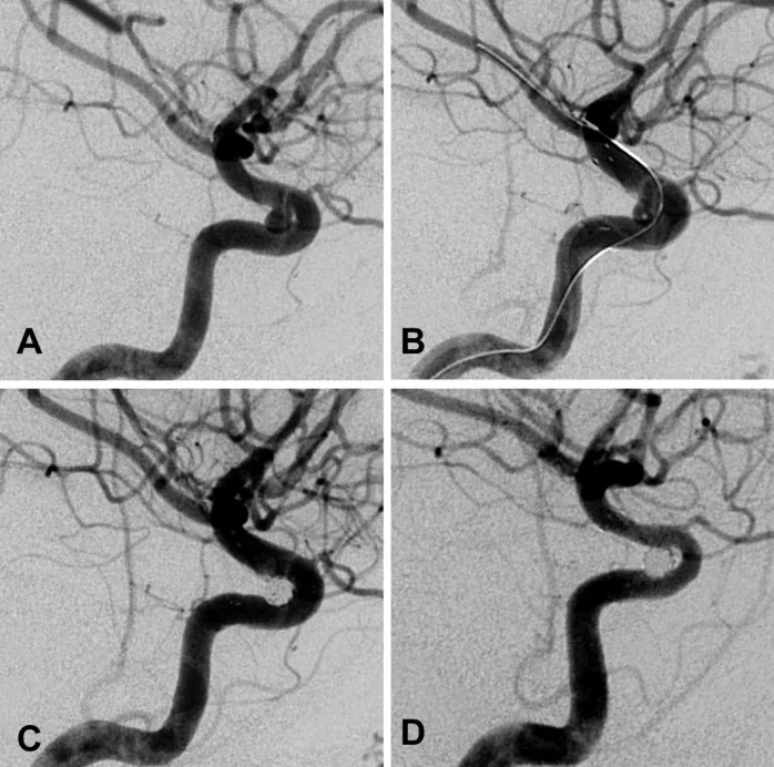
Using high-resolution three-dimensional rotational angiography, a detailed 3D X-ray imaging method, the authors measured the artery before and right after treatment and at later follow-up. The stent caused no immediate change. At follow-up, the artery inside and just beyond the stent had narrowed by an average of 0.31 to 0.41 millimeters, with the greatest narrowing (19%) near the start of the stent. This narrowing was not linked to the ischemic events (problems from reduced blood flow) that patients reported.
Longer follow-up is needed to understand its cause and importance.
A plain-language summary for patients and families. It is not medical advice, and individual results vary.
Abstract
Objective
To determine the effect of Neuroform stent (Boston Scientific/Target, Fremont, CA) deployment on parent vessel lumen and detect in-stent changes in patients harboring wide-necked intracranial aneurysms treated with the stent-coil technique.
Methods
Parent vessel dimensions were quantified before and after the procedure and at intermediate follow-up examinations by use of high-resolution three-dimensional rotational angiography. By use of shaded surface segmentation of the acquired volume, measurements of the parent vessel proximal to the stent (Point A), at three points within the stented vascular segment (Points B, C, and D), and distal to the stent (Point E) at each study time were compared by use of paired t tests. Correlation between degree of in-stent stenosis and reported ischemic events was estimated by use of a linear regression model.
Results
Stent and coil deployment had no immediate effect on parent vessel dimensions. At angiographic follow-up, there was no significant change in vessel size proximal to the stent. Within the stent and distal to it, however, there was a statistically significant 0.31- to 0.41-mm reduction in average diameter (P < 0.001, P < 0.011, P < 0.003, and P < 0.014 for Points B, C, D, and E, respectively). The highest degree of stenosis occurred at Point B, with an average decrease in cross sectional surface area of 2.4 mm (P < 0.001), corresponding to a 19% stenosis and 52% estimated increase in focal hemodynamic resistance by Poiseuille's law. No clinical correlation was noted with the degree of in-stent stenosis.
Conclusion
Intracranial stenting using a soft self-expanding stent without angioplasty induced a statistically, but not clinically, significant decrease in cross sectional area. Further research and longer-term follow-up are needed to elucidate the mechanism and clinical importance of this response.
Abstract as indexed in PubMed (PMID 16331171). Copyright the publisher.
Cite this paper
Hoit DA, Malek AM. Three-dimensional rotational angiographic detection of in-stent stenosis in wide-necked aneurysms treated with a self-expanding intracranial stent. Neurosurgery 57(6):1228-36; discussion 1228-36. 2005. doi:10.1227/01.neu.0000186026.12709.bc PMID: 16331171.