2014 · Devices & clinical
Use of cone-beam computed tomography angiography in planning for gamma knife radiosurgery for arteriovenous malformations: a case series and early report
Safain MG, Rahal JP, Raval A, Rivard MJ, Mignano JE, Wu JK, Malek AM.
Neurosurgery 74(6):682-95; discussion 695-6, 2014.
PMID 24584136 · DOI 10.1227/NEU.0000000000000331 · PMCID PMC4028381
In plain language
An arteriovenous malformation (AVM) is a tangle of abnormal blood vessels in the brain where arteries connect directly to veins. Gamma Knife radiosurgery treats AVMs with focused beams of radiation, but it works best when the entire tangle, called the nidus, is targeted while sparing healthy brain, so it depends on sharp, accurate images.
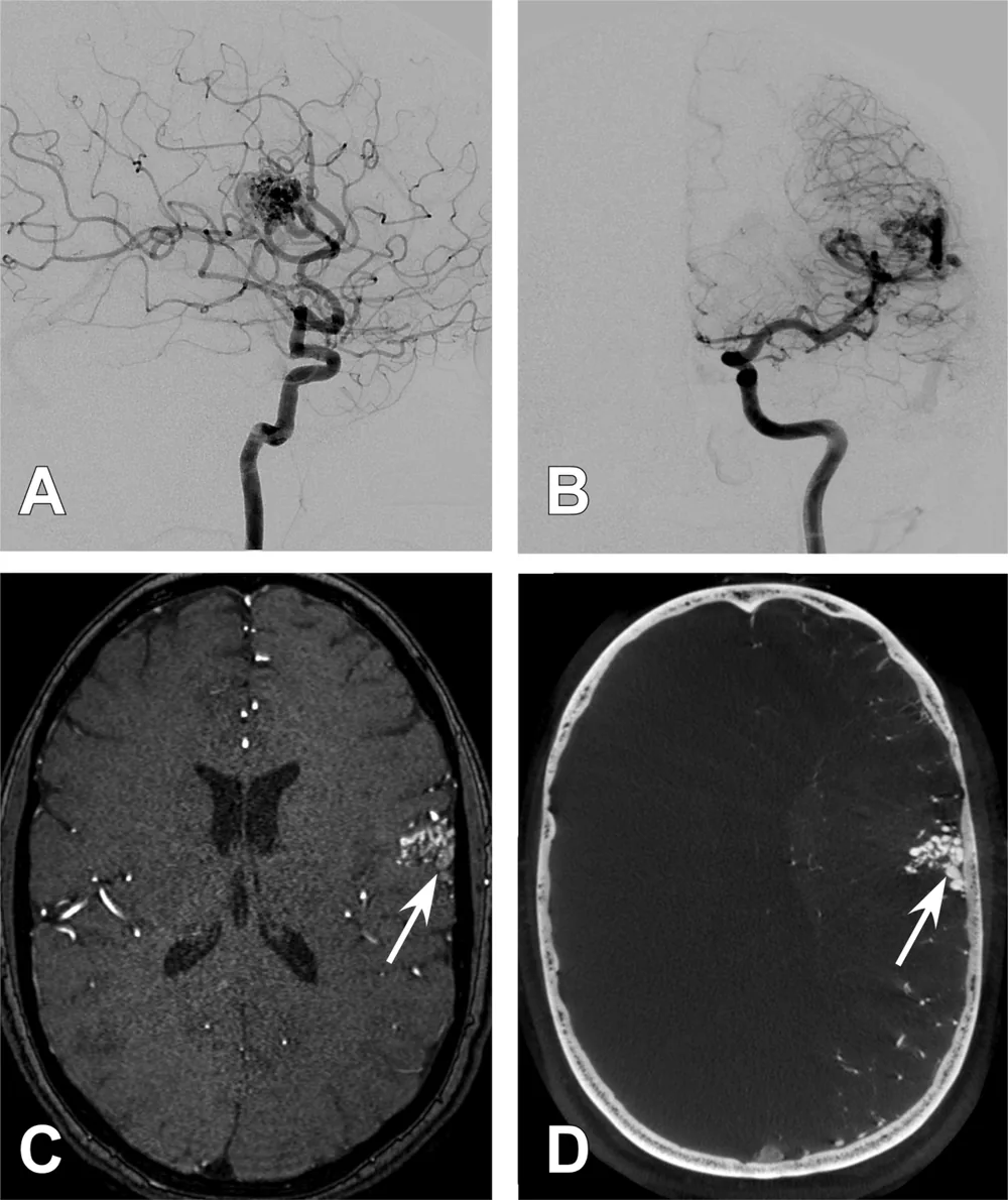
From June 2009 to June 2013, the team used cone-beam CT angiography, a very high-resolution 3D X-ray scan of the blood vessels taken with the angiography machine, to plan treatment in 22 consecutive patients. The images showed the fine structure of the nidus in detail. The average radiation dose at the edge of the AVM was 15.6 Gy. No patient had bleeding related to treatment, and at early follow-up (16 months on average), 84% of patients had an AVM that was shrinking or gone.
The authors conclude that this imaging approach is practical and useful for radiosurgery planning.
A plain-language summary for patients and families. It is not medical advice, and individual results vary.
Abstract
Background
The effectiveness of Gamma Knife radiosurgery (GKR) for cerebral arteriovenous malformations (AVMs) is predicated on inclusion of the entire nidus while excluding normal tissue. As such, GKR may be limited by the resolution and accuracy of the imaging modality used in targeting.
Objective
We present the first case series to demonstrate the feasibility of using ultrahigh-resolution C-arm cone-beam computed tomography angiography (CBCT-A) in AVM targeting.
Methods
From June 2009 to June 2013, CBCT-A was used for targeting of all patients with AVMs treated with GKR at our institution. Patients underwent Leksell stereotactic head frame placement followed by catheter-based biplane 2-dimensional digital subtraction angiography, 3-dimensional rotational angiography, as well as CBCT-A. The CBCT-A dataset was used for stereotactic planning for GKR. Patients were followed at 1, 3, 6, and 12 months and then annually thereafter.
Results
CBCT-A-based targeting was used in 22 consecutive patients. CBCT-A provided detailed spatial resolution and sensitivity of nidal angioarchitecture enabling treatment. The average radiation dose to the margin of the AVM nidus corresponding to the 50% isodose line was 15.6 Gy. No patient had treatment-associated hemorrhage. At early follow-up (mean, 16 months), 84% of patients had a decreasing or obliterated AVM nidus.
Conclusion
CBCT-A-guided radiosurgery is feasible and useful because it provides sufficient detailed resolution and sensitivity for imaging brain AVMs.
Abstract as indexed in PubMed (PMID 24584136). Copyright the publisher.
Cite this paper
Safain MG, Rahal JP, Raval A, Rivard MJ, Mignano JE, Wu JK, Malek AM. Use of cone-beam computed tomography angiography in planning for gamma knife radiosurgery for arteriovenous malformations: a case series and early report. Neurosurgery 74(6):682-95; discussion 695-6. 2014. doi:10.1227/NEU.0000000000000331 PMID: 24584136.