2010 · Hemodynamics & imaging
Automated detection of intracranial aneurysms based on parent vessel 3D analysis
Lauric A, Miller E, Frisken S, Malek AM.
Med Image Anal 14(2):149-59, 2010.
PMID 20004607 · DOI 10.1016/j.media.2009.10.005
In plain language
Most bleeding around the brain that is not caused by injury, called subarachnoid hemorrhage, comes from a ruptured aneurysm, a weak bulge in an artery wall. Spotting aneurysms on scans is important, yet some are missed.
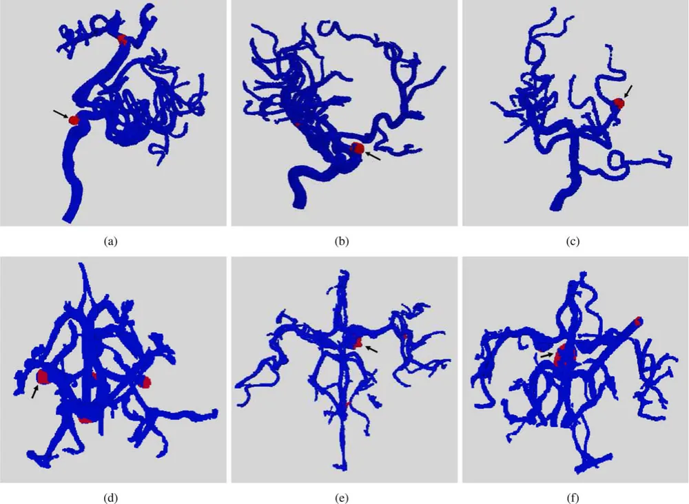
This study presents a computer method that automatically flags possible aneurysms on 3D images of the brain's blood vessels. It traces each vessel and uses a shape measure called the writhe number to find spots that do not look like a normal tube. Tested on two kinds of vessel imaging, 3D rotational angiography and CT angiography, it found 100% of aneurysms, with an average of 0.66 false alarms per study on the first and 5.36 on the second.
The tool is designed to assist doctors reading the scans.
A plain-language summary for patients and families. It is not medical advice, and individual results vary.
Abstract
The detection of brain aneurysms plays a key role in reducing the incidence of intracranial subarachnoid hemorrhage (SAH) which carries a high rate of morbidity and mortality. The majority of non-traumatic SAH cases is caused by ruptured intracranial aneurysms and accurate detection can decrease a significant proportion of misdiagnosed cases. A scheme for automated detection of intracranial aneurysms is proposed in this study. Applied to the segmented cerebral vasculature, the method detects aneurysms as suspect regions on the vascular tree, and is designed to assist diagnosticians with their interpretations and thus reduce missed detections. In the current approach, the vessels are segmented and their medial axis is computed. Small regions along the vessels are inspected and the writhe number is introduced as a new surface descriptor to quantify how closely any given region approximates a tubular structure. Aneurysms are detected as non-tubular regions of the vascular tree. The geometric assumptions underlying the approach are investigated analytically and validated experimentally. The method is tested on 3D-rotational angiography (3D-RA) and computed tomography angiography (CTA). In our experiments, 100% sensitivity was achieved with average false positives rates of 0.66 per study on 3D-RA data and 5.36 false positive rates per study on CTA data.
Abstract as indexed in PubMed (PMID 20004607). Copyright the publisher.
Cite this paper
Lauric A, Miller E, Frisken S, Malek AM. Automated detection of intracranial aneurysms based on parent vessel 3D analysis. Med Image Anal 14(2):149-59. 2010. doi:10.1016/j.media.2009.10.005 PMID: 20004607.