2010 · Hemodynamics & imaging
Spatio-temporal data fusion for 3D+T image reconstruction in cerebral angiography
Copeland AD, Mangoubi RS, Desai MN, Mitter SK, Malek AM.
IEEE Trans Med Imaging 29(6):1238-51, 2010.
PMID 20172817 · DOI 10.1109/TMI.2009.2039645
In plain language
Angiography uses X-rays and injected contrast dye to show blood vessels, but doctors usually see either a still 3D picture or flat 2D movies of the flow. This study describes a mathematical method that combines a single 3D model with two sets of 2D X-ray movies to build a 3D movie of blood moving through the brain's arteries.
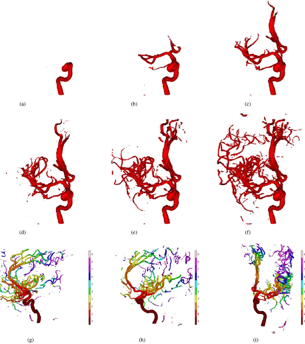
The results can be shown in several ways, including 3D surfaces color-coded by when the contrast arrives. The method was tested on computer-simulated data, and its image quality was measured.
The authors suggest it could help doctors find problems such as clots, narrowings and aneurysms more quickly during procedures, with less X-ray exposure.
A plain-language summary for patients and families. It is not medical advice, and individual results vary.
Abstract
This paper provides a framework for generating high resolution time sequences of 3D images that show the dynamics of cerebral blood flow. These sequences have the potential to allow image feedback during medical procedures that facilitate the detection and observation of pathological abnormalities such as stenoses, aneurysms, and blood clots. The 3D time series is constructed by fusing a single static 3D model with two time sequences of 2D projections of the same imaged region. The fusion process utilizes a variational approach that constrains the volumes to have both smoothly varying regions separated by edges and sparse regions of nonzero support. The variational problem is solved using a modified version of the Gauss-Seidel algorithm that exploits the spatio-temporal structure of the angiography problem. The 3D time series results are visualized using time series of isosurfaces, synthetic X-rays from arbitrary perspectives or poses, and 3D surfaces that show arrival times of the contrasted blood front using color coding. The derived visualizations provide physicians with a previously unavailable wealth of information that can lead to safer procedures, including quicker localization of flow altering abnormalities such as blood clots, and lower procedural X-ray exposure. Quantitative SNR and other performance analysis of the algorithm on computational phantom data are also presented.
Abstract as indexed in PubMed (PMID 20172817). Copyright the publisher.
Cite this paper
Copeland AD, Mangoubi RS, Desai MN, Mitter SK, Malek AM. Spatio-temporal data fusion for 3D+T image reconstruction in cerebral angiography. IEEE Trans Med Imaging 29(6):1238-51. 2010. doi:10.1109/TMI.2009.2039645 PMID: 20172817.