2008 · Hemodynamics & imaging
Estimation of wall shear stress dynamic fluctuations in intracranial atherosclerotic lesions using computational fluid dynamics
Schirmer CM, Malek AM.
Neurosurgery 63(2):326-34; discussion 334-5, 2008.
PMID 18797363 · DOI 10.1227/01.NEU.0000313119.73941.9E
In plain language
Intracranial stenosis is a narrowing of an artery inside the skull, and it can cause stroke by reducing blood flow or by shedding clots. The authors used computational fluid dynamics, computer simulation of blood flow, to study wall shear stress, the rubbing force of flowing blood on the vessel wall, which strongly affects how endothelial cells (the cells that line blood vessels) behave.
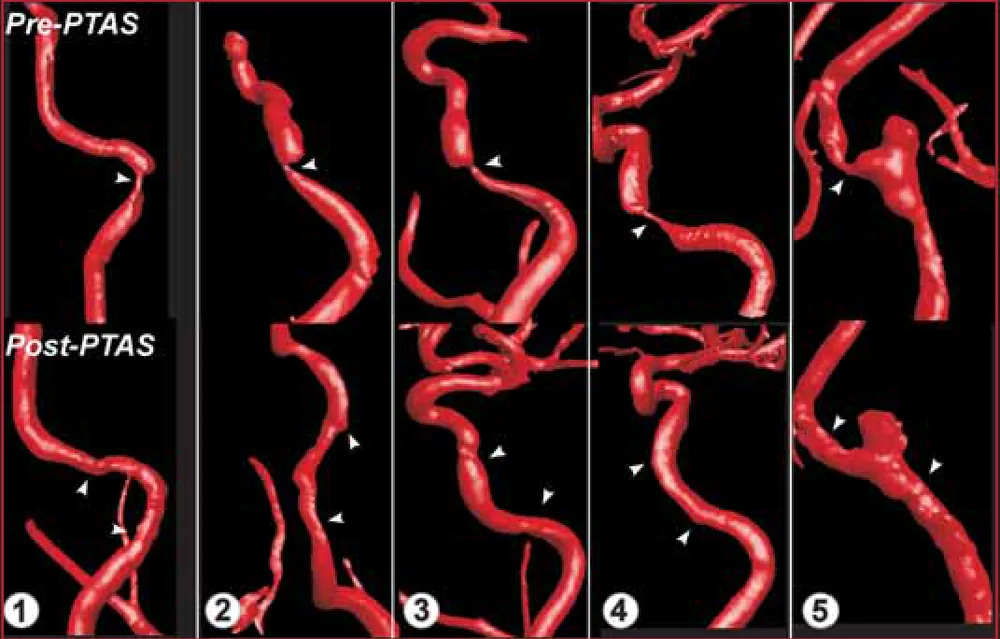
Using detailed 3D angiography images, they modeled five narrowed arteries before and after balloon angioplasty and stenting. Inside the narrowing, the force reached an average of 1870 dyn/cm and rapidly swung in direction and strength over each heartbeat, a pattern not seen in healthy segments. Treatment brought these extreme forces back toward normal.
The study reveals a harsh flow environment that may help explain how these narrowings develop.
A plain-language summary for patients and families. It is not medical advice, and individual results vary.
Abstract
Objective
Intracranial stenosis (IS) is associated with significant morbidity and mortality from hypoperfusion and thromboembolism. We used computational fluid dynamic methods to analyze luminal patterns of wall shear stress (WSS), a known critical modulator of endothelial function, within patient-based IS lesions undergoing percutaneous angioplasty and stenting.
Methods
High-resolution three-dimensional rotational angiographic data sets were reconstructed to yield a fine-resolution computational mesh allowing application of pulsatile computational fluid dynamic analysis with a non-Newtonian realistic model of blood. WSS and its gradient were analyzed spatiotemporally in five IS lesions before and after percutaneous angioplasty and stenting.
Results
WSS within the stenosis reached average shear magnitudes of 1870 +/- 783 dyn/cm with rapidly reversing direction to oscillating low values in the recirculation zone. WSS vectors revealed complex dynamic directional and amplitude oscillations not seen in healthy segments with time-dependent convergence and divergence strips sweeping back and forth across the lesion during the cardiac cycle. These areas also underwent extreme temporal WSS oscillation of 2052 +/- 909 dyn/cm over a short time interval. The endothelial mechanotransductive response to such extreme WSS magnitudes and gradients, which were normalized by percutaneous angioplasty and stenting in the current study, remains undefined.
Conclusion
Computational fluid dynamic analysis of IS has uncovered a complex and hostile microhemodynamic environment characterized by wide and rapid shear variations in time and space. Characterization of the mechanical forces acting on the wall can help in determining the molecular transduction response of the luminal endothelium to these extreme stresses and may lead to better understanding of the hemodynamic contribution to stenosis pathophysiology.
Abstract as indexed in PubMed (PMID 18797363). Copyright the publisher.
Cite this paper
Schirmer CM, Malek AM. Estimation of wall shear stress dynamic fluctuations in intracranial atherosclerotic lesions using computational fluid dynamics. Neurosurgery 63(2):326-34; discussion 334-5. 2008. doi:10.1227/01.NEU.0000313119.73941.9E PMID: 18797363.