2026 · Hemodynamics & imaging
A Novel Application of Radiomics Analysis for Computational Fluid Dynamics Evaluation of Cerebral Aneurysms
Lauric A, Malek AM.
World Neurosurg 205:124602, 2026.
PMID 41173099 · DOI 10.1016/j.wneu.2025.124602
Narrated summary
Figures from the open access article (CC BY 4.0).
Read the transcript
Here's a short summary of our recent study on blood flow inside brain aneurysms, with my longtime colleague Dr. Alexandra Lauric at Tufts Medical Center. I'm Adel Malek. When a brain aneurysm is found before it bleeds, the hardest question is whether it is likely to rupture. Size and shape help, but they don't tell the whole story. The blood flow inside the aneurysm may hold more clues. Using computational fluid dynamics, we simulated blood flow inside 68 aneurysms built from three-dimensional angiograms.
Twenty-nine of them had ruptured. The new step was to treat the flow like an image. We sampled the speed of blood on an even grid inside each aneurysm, turned it into a grayscale volume, and analyzed it with radiomics: software that measures texture and patterns, as it would in a medical scan. Here are two aneurysms side by side. In the ruptured one, slow flow fills most of the dome, with fast flow only near the neck.
In the unruptured one, faster flow reaches farther into the dome. Across 93 radiomics features, ruptured aneurysms had more uniform, lower-velocity flow inside the dome, with fewer high-speed regions and less variation in texture. Combining a few of these features, a model distinguished ruptured from unruptured aneurysms with an area under the curve of 0.90, with 86 percent sensitivity and 85 percent specificity. Radiomics, a tool built for medical images, can also read the patterns of blood flow.
This opens new ways to study why aneurysms rupture, and may one day help assess risk.
In plain language
Radiomics is a computer method that measures texture and patterns in images. Because angiograms of aneurysms look fairly uniform inside, radiomics had mainly been used to describe aneurysm shape. This study instead applied it to maps of blood speed inside the aneurysm, calculated with computer simulations of blood flow.
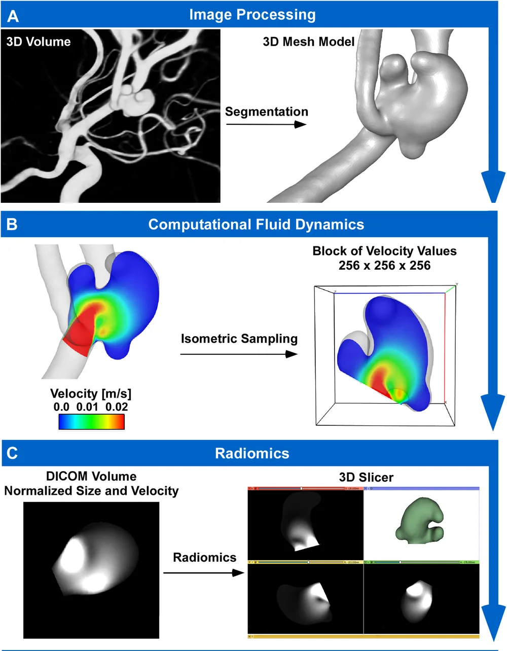
In 68 aneurysms, 29 of which had ruptured, the speed maps were converted into grayscale images and 93 features were measured. Ruptured aneurysms had more uniform patterns, less variation, fewer high-speed areas and more clustering of slow-flow regions. A combined model separated ruptured from unruptured aneurysms with an area under the curve of 0.90 (1.0 would be perfect).
This approach opens new directions for studying aneurysm rupture.
A plain-language summary for patients and families. It is not medical advice, and individual results vary.
Abstract
Background
PyRadiomics is a recently introduced automatic imaging tool for evaluating intensity-based texture descriptors and shape morphology. Due to intensity homogeneity in contrast-enhanced rotational angiography, radiomics analysis for intracranial aneurysms was previously limited to morphological evaluation. We propose a novel use of radiomics analysis to assess aneurysm intra-dome velocity distributions derived from computational fluid dynamics (CFD) simulations.
Methods
Catheter 3D rotational angiographic volumes from 68 aneurysms (29 ruptured) were analyzed. Steady-state CFD simulations were performed on segmented models, and intra-dome velocity distributions were extracted using mesh-independent isometric sampling. Velocity volumes were normalized for size and magnitude, converted to grayscale DICOM, and analyzed to extract 93 histogram and texture radiomics features. Uni- and multivariate analyses assessed these features for rupture status discrimination.
Results
Radiomics analysis showed that ruptured aneurysms had significantly lower energy (P = 0.005) and entropy (P = 0.004), but higher skewness (P = 0.001), kurtosis (P < 0.001), and uniformity (P = 0.001) than unruptured ones. Texture analysis revealed lower contrast (P = 0.02) and gray-level variance (P = 0.02), but higher strength (P = 0.04). Ruptured aneurysms also showed greater clustering of low gray-level values, reflected by higher LowGrayLevelEmphasis (P = 0.001) and LargeAreaLowGrayLevelEmphasis (P = 0.01). Multivariate analysis achieved an AUC of 0.90 (sensitivity 0.86, specificity 0.85).
Conclusion
Automatic histogram and texture analysis of intra-dome velocity using radiomics showed strong rupture status discrimination. Ruptured aneurysms exhibited pattern uniformity with reduced gray-level variability, and fewer high-intensity areas. This novel approach to CFD volumetric evaluation, extending radiomics use beyond angiographic analysis, provides insights into rupture characteristics, and opens novel and promising avenues of future research.
Abstract as indexed in PubMed (PMID 41173099). Copyright the publisher.
Cite this paper
Lauric A, Malek AM. A Novel Application of Radiomics Analysis for Computational Fluid Dynamics Evaluation of Cerebral Aneurysms. World Neurosurg 205:124602. 2026. doi:10.1016/j.wneu.2025.124602 PMID: 41173099.