2026 · Hemodynamics & imaging
A novel application of radiomics analysis for the evaluation of intradome transient velocity in cerebral aneurysms
Lauric A, Malek AM.
J Neurosurg 144(5):1044-1053, 2026.
PMID 41616305 · DOI 10.3171/2025.9.JNS251068
In plain language
Blood flow inside an aneurysm changes throughout each heartbeat, but there are few ways to measure those changes in detail. This study used radiomics, a method that analyzes texture and pattern in images, on blood speed data from computer flow simulations over a full heartbeat.
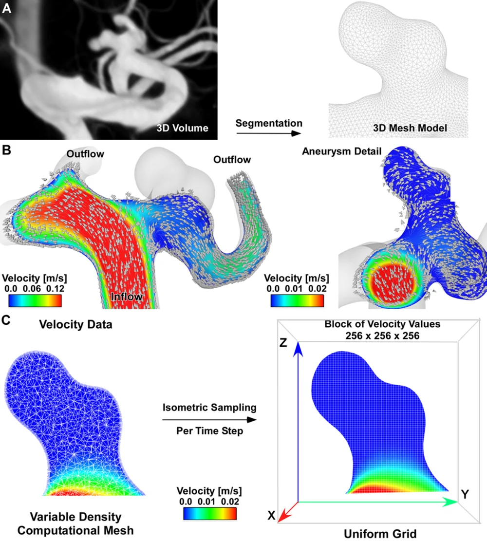
In 75 aneurysms, 33 of which had ruptured, the speed data at each moment were flattened into images and analyzed. Ruptured aneurysms were dominated by slow-flow regions with consistent textures, but also had small pockets of fast flow, suggesting uneven flow. A combined model separated ruptured from unruptured aneurysms with an area under the curve of 0.88 (1.0 would be perfect).
The method extends radiomics to flow changes across the heartbeat and may guide future research on rupture risk.
A plain-language summary for patients and families. It is not medical advice, and individual results vary.
Abstract
Objective
Volumetric hemodynamic analysis has previously demonstrated robustness to surface irregularities and noise; however, quantitative evaluation methods remain limited. The authors introduce a novel approach to assess intradome velocity distributions in intracranial aneurysms by applying radiomics texture and pattern analysis to time-step data derived from transient computational fluid dynamics (CFD) simulations.
Methods
Angiographic volumes from catheter 3D rotational angiography were available for 75 aneurysms (33 ruptured). Transient CFD simulations were performed on aneurysm models over an entire cardiac cycle. Intradome volumetric velocity distributions were exported for consecutive time points, followed by maximum intensity projection (MIP) to 2D images and logarithmic transformation to enhance discrimination analysis at the intensity extrema. Two-dimensional images representing consecutive time points were used as input to radiomics as 3D DICOM volumes. Univariate and multivariate analyses assessed rupture status discrimination accuracy of 93 histogram, texture, and pattern radiomics features.
Results
Radiomics analysis of MIP velocity texture and pattern revealed that, compared with unruptured aneurysms, ruptured aneurysms have lower intensity patterns (higher energy and total energy of logarithmic values), reflecting dominant low-velocity regions. These regions exhibit consistent intensity textures (higher correlation and inverse difference, with lower cluster shade and difference entropy) with recurrent patterns of similar intensities (higher maximum probability and long-run emphasis). Localized high-velocity runs are also present (higher high gray-level run emphasis) in ruptured aneurysms, suggesting a heterogeneous flow profile. Stepwise regression multivariate analysis achieved rupture status discrimination with an area under the curve of 0.88 (sensitivity 0.91, specificity 0.74).
Conclusions
The automatic analysis of transient velocity using radiomics, incorporating pattern and texture analysis, demonstrated strong discriminating performance for rupture status. This innovative application of radiomics expands its utility beyond tumor and aneurysm shape analysis, enabling evaluation of velocity changes throughout the entire cardiac cycle. It provides valuable insights into rupture characteristics and paves the way for exploring future research possibilities.
Abstract as indexed in PubMed (PMID 41616305). Copyright the publisher.
Cite this paper
Lauric A, Malek AM. A novel application of radiomics analysis for the evaluation of intradome transient velocity in cerebral aneurysms. J Neurosurg 144(5):1044-1053. 2026. doi:10.3171/2025.9.JNS251068 PMID: 41616305.